


Why Having "Perfect 20/20 Vision" Can Mask a Hidden Blood Leak Inside Your Eye
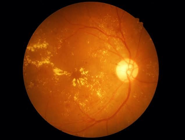
The Dangerous Assumption: "My Vision is Fine, So My Eyes Are Safe"
It is one of the most frequent—and risky—misconceptions heard in primary care and optometry clinics throughout Downtown Houston, Sugar Land, and Webster. A patient is diagnosed with Type 2 diabetes or prediabetes. They manage their diet, take their medication, and check their A1C. But because they can still read the television screen clearly and drive without glasses, they skip their annual medical eye evaluation.
They assume that if diabetes were damaging their eyes, they would feel pain or notice blurry vision.
At Evolutionary Eye Care, we want to deliver a direct, crucial medical truth: early diabetic eye disease produces zero pain and zero early vision symptoms.
By the time you notice floating spots, distorted straight lines, or a sudden drop in visual clarity, blood vessels inside your retina may have been silently leaking fluid for months. In 2026, catching these microscopic capillary shifts early is the single most effective way to prevent permanent sight loss.
The Glucose Erosion: How Sugar Damages the Retinal Micro-Capillaries
Your retina—the light-sensing tissue lining the back wall of your eye—has one of the highest metabolic demands of any tissue in the human body. To feed this intense workload, it relies on an ultra-dense, microscopic network of tiny blood vessels (capillaries).
When blood glucose levels remain elevated over time, or fluctuate wildly throughout the day, excess sugar molecules physically weaken the structural walls of these fragile micro-capillaries.
[Elevated Blood Glucose] -> [Pericyte Cell Loss in Capillaries] -> [Microaneurysms Form (Ballooning)] -> [Fluid Leaks into Retina] -> [Diabetic Macular Edema (DME)]
This structural erosion progresses through distinct pathological stages:
| Stage of Diabetic Retinopathy | What Happens Inside the Eye | Symptoms Experienced |
| Mild Non-Proliferative (NPDR) | Microscopic balloon-like swellings (microaneurysms) form in capillary walls. | None. Vision remains crisp and 20/20. |
| Moderate to Severe NPDR | Capillaries become blocked, depriving areas of the retina of oxygen-rich blood. | Subtle loss of night vision or contrast sensitivity. |
| Diabetic Macular Edema (DME) | Leaking blood and fluid pool directly inside the macula (central vision zone). | Central blur, washed-out colors, or straight lines appearing wavy. |
| Proliferative (PDR) | Oxygen-starved retina grows fragile new blood vessels that break and bleed. | Sudden shower of dark floaters, cobwebs, or complete visual blockage. |
The 2026 Interception Track: 3D OCT & Non-Invasive Vascular Mapping
Waiting for visual blur before scheduling an eye exam means waiting until structural fluid damage has already reached your central macula. At our Webster, Houston, and Sugar Land offices, we utilize advanced medical retinal imaging to intercept diabetic vascular changes long before they touch your vision:
1. High-Definition Spectral-Domain OCT
We capture micro-thin optical cross-sections of your retinal layers down to the micrometer. This non-invasive light scan allows us to spot microscopic pockets of fluid pooling beneath the macula, measuring swelling volume before it impacts visual acuity.
2. OCT Angiography (OCT-A)
Using laser-derived flow tracking, we map blood movement through your retinal capillaries in real-time without injecting intravenous dye. This enables us to detect areas of capillary non-perfusion (oxygen starvation) and microaneurysms early.
3. Collaborative Systemic Co-Management
We don't manage your eyes in isolation. Following your medical exam, we send a detailed structural retinal report to your endocrinologist or primary care physician. Tracking micro-vascular changes in the eye provides a clear window into how well your systemic micro-vascular health (including your kidney capillaries) is holding up.
The Corneal Nerves and Neurotrophic Dry Eye
Diabetic vascular erosion isn't limited to the back of the eye. Sustained elevated blood sugar also causes diabetic neuropathy in the delicate nerve fibers feeding your cornea—the front surface of your eye.
When corneal nerves lose sensitivity, your brain stops receiving signals to blink and produce natural tears. This leads to an aggressive form of Neurotrophic Dry Eye, where the corneal surface develops microscopic dry spots without triggering typical pain signals.
At our Dry Eye Center of Excellence Houston, we treat this surface vulnerability directly. We deploy non-invasive Low-Level Light Therapy (LLLT) at our Sugar Land and Houston locations to stimulate cellular ATP, unblock Meibomian glands, and restore a protective oil seal over your tears. Maintaining a smooth, lubricated corneal surface ensures diagnostic retinal scans remain precise.
Whether you are preserving your visual stamina for high-demand corporate careers, optimizing visual reflexes for Sports Vision & Performance, or fitting specialized optics like Scleral Lenses or Adult Ortho-K, a healthy vascular baseline protects your sight.
Medical Eye Exam vs. Routine Vision Test
A standard retail refraction checkup measures your basic eyeglass prescription, but it cannot evaluate retinal capillary integrity or measure sub-retinal fluid accumulation.
Our comprehensive Medical Eye Exam protocol includes widefield digital retinal imaging, high-definition 3D OCT macular scans, and corneal sensitivity assessments.
Conclusion: Protect Your Visual Capital
Having diabetes or prediabetes does not mean vision loss is inevitable. Early diabetic retinopathy and macular edema are manageable medical conditions when caught in time. Do not wait for visual blur to tell you there is a problem. By combining non-invasive 3D retinal mapping, vascular OCT tracking, and advanced surface light therapies, we can detect early capillary shifts, protect your macula, and safeguard your vision for a lifetime of health and independence.


WHERE TO FIND US Our Locations

© 2026 Evolutionary Eye Care . All Rights Reserved. Accessibility Statement - Privacy Policy - Sitemap
Powered by:

