


Why Hearing a "Whooshing" Rhythm in Your Ears and Sudden Visual Blackouts Aren't Just Migraines
The Whooshing Rhythm: When Headaches Come with Visual Blackouts
It is a deeply alarming combination of symptoms experienced by young adults and working professionals across Downtown Houston, Sugar Land, and Webster. You bend down to tie your shoe or stand up quickly from your desk, and your vision momentarily goes pitch black in one or both eyes for a few seconds (transient visual obscurations).
Accompanying these visual blackouts is a persistent, heavy headache behind your eyes that feels worse when lying down, along with a strange "whooshing" or thumping sound in your ears synchronized with your heartbeat (pulsatile tinnitus).
When you consult a primary doctor, these episodes are often initially brushed off as atypical migraines, sinus pressure, or dehydration.
At Evolutionary Eye Care, we evaluate these symptoms with immediate neuro-optometric urgency. Experiencing visual blackouts alongside heart-rate-synchronized ear roaring indicates that elevated fluid pressure inside your skull is physically crushing your optic nerves—a medical condition known as Idiopathic Intracranial Hypertension (IIH) or Pseudotumor Cerebri.
The Fluid Squeeze: How Brain Pressure Causes Papilledema
Your brain and spinal cord are bathed in protective cerebrospinal fluid (CSF). Under normal conditions, your body produces and reabsorbs CSF at a continuous, balanced rate.
In Idiopathic Intracranial Hypertension, the body fails to drain CSF properly, leading to a buildup of intracranial pressure without a brain tumor present.
[CSF Fluid Drainage Backup] -> [Elevated Intracranial Pressure] -> [Pressure Travels Down Optic Nerve Sheath] -> [Optic Disc Swelling (Papilledema)]
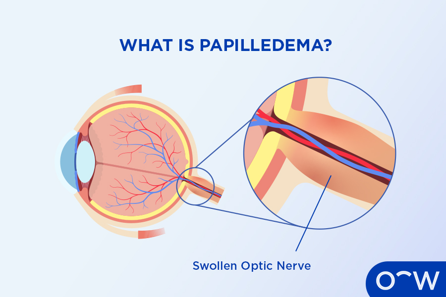
Because the optic nerve connects directly to the brain, high fluid pressure travels along the nerve sheath, squeezing the delicate nerve fibers where they enter the back of the eye. This pressure-induced optic nerve swelling is called Papilledema. As the nerve swells, blood flow stalls, causing brief visual blackouts and threatening permanent optic nerve atrophy if left unmanaged.
IIH Symptoms vs. Standard Tension Migraines
| Diagnostic Marker | Standard Tension / Migraine Headache | Idiopathic Intracranial Hypertension (IIH) |
|---|---|---|
| Headache Timing | Triggered by stress or light; improves with rest. | Worse upon waking or lying flat; worsens when coughing or bending over. |
| Auditory Signs | Sensitivity to loud noise (phonophobia). | Pulsatile Tinnitus: Hearing a "whooshing" sound tuned to your heartbeat. |
| Visual Changes | Visual auras (jagged lines) lasting 20-30 minutes. | Transient Visual Obscurations: Sudden 2-5 second visual blackouts when changing posture. |
| Optic Nerve State | Optic nerve margins look flat and healthy. | Papilledema: Bilateral, severe optic nerve elevation and swelling. |
The 2026 Neuro-Diagnostic & Co-Management Protocol
Catching optic nerve swelling early prevents permanent visual field loss. As a specialized Neuro-Optometric Center of Excellence, we deploy an advanced diagnostic protocol across our Webster, Houston, and Sugar Land clinics:
1. High-Definition 3D Optical Coherence Tomography (OCT)
We capture micro-thin cross-sectional scans of your optic nerve head down to the micrometer. OCT mapping quantifies retinal nerve fiber layer (RNFL) swelling, providing an objective baseline to monitor changes in fluid pressure over time.
2. Automated Threshold Visual Field Mapping (30-2 / 60-4)
Papilledema selectively damages peripheral vision before touching central acuity. We run precision visual field mapping to detect subtle blind spot enlargement and peripheral field loss caused by nerve compression.
3. Interdisciplinary Neurology Co-Management
We collaborate directly with top neurologists and neuro-ophthalmologists in Houston to coordinate brain MRIs and MRVs (ruling out venous sinus thrombosis) and guide therapeutic lumbar punctures or medical therapies (such as acetazolamide) to lower intracranial fluid pressure.
Surface Relief and Dry Eye Intersect
High intracranial pressure and the medications used to manage IIH often cause systemic dehydration, degrading tear film quality and triggering severe Evaporative Dry Eye.
At our Dry Eye Center of Excellence Houston, we protect your corneal surface during neuro-rehabilitation. We deploy non-invasive Low-Level Light Therapy (LLLT) at our Sugar Land and Webster locations to clear congested Meibomian glands, cool lid inflammation, and maintain a protective oil seal over your tears—keeping your ocular surface smooth while your optic nerves recover.
Whether you are preserving visual stamina for corporate performance, fine-tuning tracking for Sports Vision & Performance, or integrating care with Scleral Lenses or Adult Ortho-K, protecting the optic nerve pathway is paramount.
Medical Eye Exam vs. Routine Vision Test
Papilledema and IIH cannot be detected through a basic storefront 20/20 vision chart. Reading letters on a distance wall test proves your central lens works under ideal conditions, completely missing severe peripheral optic nerve swelling.
Our comprehensive Medical Eye Exam for intracranial pressure includes dilated optic nerve evaluations, high-definition 3D OCT RNFL scans, automated visual field testing, and fundus photography.
Conclusion: Protect Your Optic Nerves from Pressure
Sudden visual blackouts when standing and a whooshing sound in your ears are clear cues that your optic nerves are under physical pressure. Idiopathic Intracranial Hypertension and Papilledema are serious, manageable neuro-optometric conditions, and early diagnostic imaging prevents permanent sight loss. By combining 3D OCT nerve mapping, automated visual field tracking, and collaborative neurology co-management, we can detect optic nerve swelling, track fluid resolution, and safeguard your vision for life.


WHERE TO FIND US Our Locations

© 2026 Evolutionary Eye Care . All Rights Reserved. Accessibility Statement - Privacy Policy - Sitemap
Powered by:

