


Why Your "Severe Astigmatism" Might Actually Be a Rare Corneal Bulge (And Why Glasses Fail)
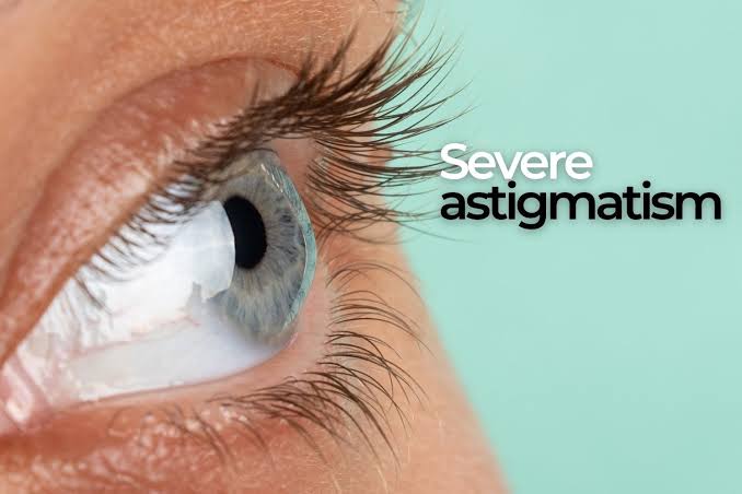
The 2026 Specialty Optics Protocol: Vaulting the Inferior Bulge
You cannot correct a warped, asymmetric PMD cornea with standard flat eyeglasses or soft contacts. Attempting to rest a rigid lens directly on an inferior corneal bulge risks surface scarring.
As a specialized Specialty Lens and Ocular Surface Center of Excellence, we deploy an advanced fitting track at our Webster, Houston, and Sugar Land clinics:
1. Quadrant-Specific Scleral Lenses
We design custom Scleral Lenses that rest on the firm white sclera, floating completely over the delicate inferior bulge. Because the lower portion of a PMD cornea protrudes significantly, we utilize quadrant-specific technology—customizing the landing curve of each 90-degree quadrant of the lens independently. The lens stays stable, never touches the thinned tissue, and holds a continuous saline bath over the eye to erase irregular astigmatism.
2. Corneal Collagen Cross-Linking (CXL) Co-Management
If 3D tomography confirms that your inferior thinning is actively progressing, we co-manage Corneal Cross-Linking (CXL) with top corneal specialists in Houston to stiffen the collagen fibers and lock the disease in place before fitting final scleral optics.
3. Surface Reset via Low-Level Light Therapy (LLLT)
Severe corneal asymmetry disrupts normal tear spreading. We deploy non-invasive Low-Level Light Therapy (LLLT) at our Sugar Land and Webster offices to unblock Meibomian glands, cool lid inflammation, and maintain a protective oil seal over your tears—ensuring maximum scleral lens wearing comfort.
Whether you are preserving visual stamina for corporate leadership, fine-tuning spatial tracking for Sports Vision & Performance, or integrating care with Adult Ortho-K, custom scleral vaulting restores optical symmetry.
Medical Eye Exam vs. Routine Vision Test
Pellucid Marginal Degeneration cannot be caught or evaluated through a basic storefront 20/20 vision chart. Reading static letters in an exam room proves your central vision works under forced conditions, completely missing inferior peripheral thinning and back-surface corneal steepening.
Our comprehensive Medical Eye Exam for irregular astigmatism includes 3D Corneal Tomography (mapping front and back elevation contours down to the micron), pachymetric thickness mapping, and high-resolution anterior segment imaging.
Conclusion: Realign Your Vision with Precision Optics
Living with worsening astigmatism and failed contact lenses is frustrating, but Pellucid Marginal Degeneration is a manageable structural condition. By capturing 3D elevation maps of your corneal architecture, utilizing custom quadrant-specific scleral optics, and maintaining a healthy tear film, we can vault over the inferior bulge, eliminate visual distortion, and restore razor-sharp clarity.


WHERE TO FIND US Our Locations

© 2026 Evolutionary Eye Care . All Rights Reserved. Accessibility Statement - Privacy Policy - Sitemap
Powered by:

